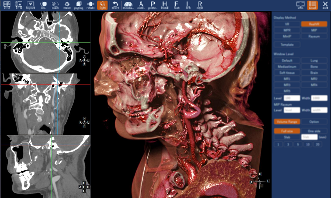
We have newly re-engineered our original 3D viewer for use in medicine and dentistry, and have completely redesigned the user interface.
Our 3D viewer system for CT and MR images, which we have been developing since 2016 for use in medicine and dentistry, has now been redesigned with a completely new user interface.
In addition, several new features and improvements have been implemented this time. We will continue enhancing the functionality of this system in the future.

The top toolbar of the 3D Viewer is designed with buttons that are easy to understand at a glance. The right side panel for manipulating MPR and volume rendering images has been redesigned to be sleeker and easier to operate.

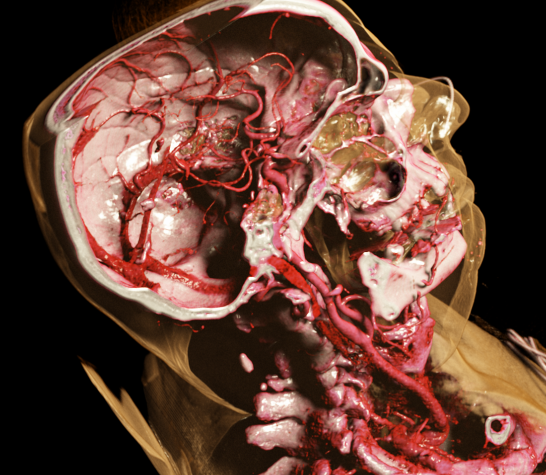
This high-impact image here is a volume-rendered image from our new 3D viewer with our photorealistic 3D rendering engine.
This image realistically depicts the anatomical structures of the arteries and each bone in the head and neck region. The image also clearly shows how the internal carotid artery connects from the bottom to the top through the skull base to arteries in the brain.


